New Mammogram Technology
Dr. Bryan GushikenRadiologist at Kapiolani Women’s Center
Where did you receive your schooling/training?
I attended the University of Southern California and medical school at UCLA. I did my residency, radiology and fellowship at the University of California San Francisco.
How long have you been in practice?
Since 1997.
What does a traditional mammogram look like?
Traditional mammography is two-dimensional. The 2-D mammogram consists of four pictures, two of each breast, that every woman should be getting annually from the age of 40. The breast is placed into position, and we compress the breast so the beam has less tissue to travel through so we can get clearer pictures. We end up with a composite image of all the tissue that the beam is going through. As radiologists, we look at the 2-D picture and sort through the overlapping tissue to find any potential cancer. That’s how it’s been for more than 20 years.
How does the new breast tomosynthesis technology work?
The biggest problem we’ve had with mammography has been overlapping tissue. When you compress the breast, if there is something hiding in there, the tissue that is above or below it can obscure it from being visualized. We have to look through the image and look at all those overlapping structures to try to find something. This is particularly problematic in dense breasts, because things can hide easily in there.
The new technology, tomosynthesis, enables us to create very thin, 1-millimeter slices through every level of the breast, and then we can scroll through and minimize overlapping tissue. If there is a cancer hiding in there, we can see it a lot more clearly, because we can look at that level and not have the tissue above and below.
Is there any downside in terms of radiation?
Radiation is a relative downside. The amount of radiation you get from a 3-D mammogram is the equivalent of getting two standard digital mammograms. The amount is so minimal that it’s still well within the FDA limit. The FDA approved this technology in 2011. Yes, it is a little more radiation, but we’re seeing things so much more clearly now, which greatly reduces the need for call-backs and rescreenings.

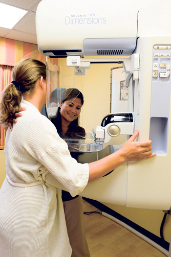
Technologist Celeste Kamihara performs a 3-D tomosynthesis mammogram on a patient during her annual screening. Nathalie Walker photos
Two-D mammograms have decreased mortality from breast cancer by 10 to 50 percent, depending on which study you look at. But 2-D mammography is far from perfect. It can miss breast cancers up to 30 percent of the time. This is where the 3-D comes in to improve that statistic.
There has been about a 30 percent increased sensitivity or pickup rate of suspicious tissue with the 3-D imaging. We got our tomosynthesis technology last August. Several times now, I have seen something that, when I looked at the regular mammogram, I could not see. It’s amazing, because on the 3-D, you can see it clearly. Who knows how long this patient would have gone before the cancer actually had shown up one year on her regular 2-D mammogram?
How often do women need a follow-up appointment with 2-D vs. 3-D?
Guidelines by the U.S. Preventative Task Force said women should consider getting a mammogram every other year. One of the things they were addressing was anxiety for patients having to go back or get so-called unnecessary biopsies. With regular screening mammo-grams, a patient gets her pictures taken and then she leaves. The radiologist reads the exam later. If they see something potentially suspicious, they call the patient back. You can imagine when the patient gets called back, it’s very anxiety-provoking, because she doesn’t know if she has something or not. Often, it turns out to be nothing. But that patient has gone through several days of anxiety.
With 3-D mammography, we visually slice through the breast. If we see something that may be a lesion or mass, we’re able to go through and see if it’s real or not. These patients may never even know that there was something initially suspected, because we were able to solve it with the 3-D data set that we already took, and they don’t get called back.
Nationally, two out of every 10 patients who get a screening mammogram get called back. With the 3-D technology, we’re getting report of a 30 to 40 percent reduction in callback.
Does insurance cover the 3-D imaging?
Not yet. Insurance is billed for the regular mammogram, and the patient is charged a separate fee for the additional 3-D part. We try to keep it relatively affordable, because we believe so much in the technology.
I think everyone should get the 3-D screening, but those who benefit most are those with dense breasts, or high-risk patients who have a family history or – just to reduce anxiety – patients who get called back all the time. With 3-D, fewer people need to come back, we find the cancers earlier and less additional screening is needed down the road.
What percentage of males get breast cancer?
Of all breast cancers, 1 percent to 2 percent are in males. Male breast cancer is a lot more deadly, because men don’t get screened every year. By the time it’s found by feeling it, it’s a lot more advanced. A lot of men ignore it; they don’t want to come in.
Anything else?
Mammogram screening can be uncomfortable, but, of course, cancer is more uncomfortable. The techs work really well with patients in terms of putting them at ease and getting them to see we’re not doing this to harm you, this is for your benefit.


